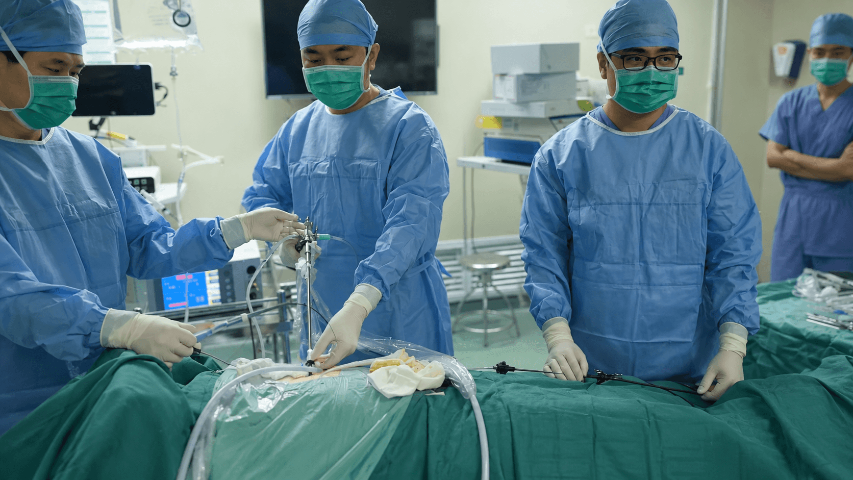
For a long time in the past, open surgical choledocholithotomy through laparotomy was the primary treatment for common bile duct stones.
With the development of gastrointestinal endoscopic technology, many patients with common bile duct stones can now undergo stone removal via ERCP. This procedure involves entering the digestive tract through the mouth and performing bile duct stone extraction, biliary drainage, stent placement, and other treatments under endoscopy.
After years of clinical research, Dr. Wang Xiaoliang from the Department of Hepatobiliary Surgery at Shanghai Pudong Hospital Affiliated to Fudan University has combined ERCP with a technique that preserves the Oddi sphincter to extract common bile duct stones through the mouth, relieving pain for a large number of patients with this condition.
What Are Common Bile Duct Stones?
Common bile duct stones refer to stones located within the common bile duct. The common bile duct is an important passageway for bile excretion. Stones here can obstruct bile flow and trigger a series of symptoms and complications.
Based on their origin, common bile duct stones can be classified into two types:
1. Primary common bile duct stones: These stones form directly within the extrahepatic bile duct. In such cases, the gallbladder may not necessarily contain stones.
2. Secondary common bile duct stones: These occur when stones from the gallbladder migrate into the common bile duct. The shape and composition of secondary stones are often similar to those of the original gallbladder stones.
Symptoms of Common Bile Duct Stones
The presentation of common bile duct stones varies significantly, with severity depending primarily on the degree of bile duct obstruction and whether biliary infection is present.
The most common symptom is upper abdominal pain, which often occurs after consuming fatty meals.
If a stone blocks the bile duct and bile cannot drain, jaundice may develop. Patients may experience yellowing of the skin and eyes, and dark-colored urine. Over time, this can progress to biliary cirrhosis.
Bile duct stones are also prone to infection. When infection occurs, patients may develop upper abdominal colic, fever, and jaundice. If the infection is severe and progresses to acute obstructive suppurative cholangitis, the condition can deteriorate rapidly, potentially leading to shock and a high mortality rate.
Additionally, biliary obstruction and inflammation caused by common bile duct stones can trigger gallstone pancreatitis. Without prompt treatment, some patients may develop severe acute pancreatitis, which is also life-threatening.
Treatment Methods for Common Bile Duct Stones
Currently, treatment for common bile duct stones falls into two main categories: surgical stone removal and endoscopic stone removal.
In the past, open choledocholithotomy was an effective treatment. However, open surgery is traumatic, recovery is slow, and some patients require a temporary bile drainage tube. For elderly, frail patients with poor underlying health, traditional surgery is poorly tolerated and may even be impossible to undertake.
With advances in gastrointestinal endoscopy, hepatobiliary surgeons have gradually developed a minimally invasive technique for removing common bile duct stones without open surgery, known as endoscopic retrograde cholangiopancreatography, or ERCP.
ERCP is a minimally invasive treatment for biliary and pancreatic diseases performed using a gastrointestinal endoscope.
The doctor inserts a duodenoscope, similar to a regular gastroscope, through the patient’s mouth. It passes through the stomach and reaches the duodenum just below. The bile and pancreatic ducts share an opening in the duodenum. Under endoscopic guidance, the doctor can perform contrast imaging of these ducts through this opening and then carry out treatments such as stone extraction, biliary drainage, and stent placement.
In simple terms, ERCP does not access the bile duct through an abdominal incision. Instead, it enters the digestive tract through the mouth and then reaches the bile duct via its natural opening in the intestine to resolve problems.
What Treatments Can ERCP Perform?
ERCP can not only remove common bile duct stones but also perform biliary drainage and biliary stent placement.
For patients with common bile duct stones, the doctor locates the biliary opening using the duodenoscope and then introduces specialized instruments into the bile duct to extract the stones.
In cases of severe biliary obstruction, significant infection, or poor patient condition, a biliary stent can be placed first to allow drainage of bile and purulent bile, relieving obstruction and controlling infection. Endoscopic stone extraction can then be scheduled once the patient’s condition improves.
ERCP Offers Advantages Over Traditional Surgery
The core advantage of ERCP is that it avoids open surgery and minimizes trauma. Compared with traditional surgery, ERCP has the following features:
No abdominal incision is required, and no endotracheal intubation anesthesia is needed. It involves minimal trauma, less pain, faster recovery, and excellent safety.
ERCP is especially valuable for elderly, frail patients who cannot tolerate surgical procedures.
It is also more suitable for patients who have previously undergone cholecystectomy and later develop residual or recurrent common bile duct stones.
As endoscopic stone retrieval instruments become increasingly refined and physician expertise continues to improve, endoscopic common bile duct stone removal has gradually replaced traditional open surgery and become one of the preferred treatments for common bile duct stones.
Which Patients Are Better Candidates for ERCP?
Elderly patients; frail patients; those with poor tolerance for surgery; patients with residual common bile duct stones after cholecystectomy; patients with recurrent common bile duct stones; and those who cannot tolerate open surgery under general anesthesia but require urgent relief of biliary obstruction.
For patients complicated by severe infection, biliary obstruction, or at risk of septic shock, ERCP can also serve as an important method for emergency decompression.
Specialized Technique by Dr. Wang Xiaoliang: Preserving the Sphincter of Oddi
During the stone extraction process, the physician uses medication to relax the sphincter of Oddi, enabling common bile duct stone removal without incising the sphincter.
The sphincter of Oddi is located at the exit where the bile and pancreatic ducts empty into the duodenum. It regulates the flow of bile and pancreatic juice and also prevents intestinal contents from refluxing into the biliary tract.
If this structure is cut, the normal barrier function of the biliary tract may be compromised. In the long term, this can potentially lead to issues such as duodenal fluid reflux, recurrent cholangitis, and recurrent common bile duct stones.
Feasible for Elderly Patients with Multiple Underlying Conditions
An 80-year-old patient with cirrhosis and ascites also presented with a large number of stones in the intrahepatic and common bile ducts. Liver resection was not an option due to cirrhosis and ascites; and given the advanced age, the patient was also unlikely to tolerate a liver transplant.
After analyzing the location and size of the stones through imaging, Dr. Wang Xiaoliang devised a step-by-step treatment plan: first, ultrasound-guided percutaneous puncture to place a 2mm diameter drainage tube in both the left and right intrahepatic bile ducts, creating a pathway between the skin surface and the intrahepatic ducts; next, dilating the tracts and using a rigid choledochoscope to enter and fragment the stones with electrohydraulic lithotripsy for removal; the common bile duct stones were then fragmented similarly, followed by efficient retrieval through the mouth via ERCP; finally, a flexible choledochoscope and an ultra-thin choledochoscope were used to clear any residual stones, meticulously inspecting each bile duct branch to confirm complete stone clearance.
The entire procedure involved no incisions and no liver resection. All stones were successfully removed, leading to restored appetite and a rapid recovery for the patient.
Can Patients Who Have Had Gastric Cancer Surgery Still Undergo ERCP?
Yes, but the difficulty increases significantly.
Consider a 78-year-old patient with a history of radical gastrectomy for cancer, who later presented with high fever, abdominal pain, and distension due to common bile duct stones. A CT scan confirmed the stones, and the patient’s blood pressure dropped to 70/40 mmHg, exhibiting classic signs of acute suppurative cholangitis and progressing to septic shock.
Given the prior radical gastrectomy and current liver metastases, the patient could not tolerate open surgery under general anesthesia. Therefore, Dr. Wang Xiaoliang’s team opted for a transoral ERCP approach.
However, the gastrointestinal anatomy is altered after a radical gastrectomy, making standard ERCP maneuvers challenging. After assessing the anatomical reconstruction based on previous surgical records, the team decided to perform the ERCP using a colonoscope.
Under colonoscopic guidance, the physician accurately identified the afferent loop of the small intestine and successfully reached the duodenal papilla. A needle-knife papillotomy was performed, a guidewire was inserted, and a plastic stent was placed, immediately allowing purulent bile to drain. Due to the patient's poor general condition, the initial strategy focused on decompression via stent placement. Once the patient's systemic status improved, elective endoscopic stone removal was scheduled. The patient’s temperature returned to normal the following day. One week later, the stones were successfully extracted during a subsequent colonoscopy-assisted ERCP procedure.
Although ERCP avoids open surgery, it remains a highly specialized form of minimally invasive endoscopic therapy. The procedure requires the physician’s careful judgment based on the patient's condition, stone location, biliary tract status, and prior surgical history.
Especially in patients with previous gastrointestinal reconstruction surgery, the complexity of ERCP increases markedly, demanding a higher level of physician experience and technical skill. Therefore, patients must seek out hepatobiliary surgeons with substantial clinical experience to perform the procedure.
Do Not Delay Treatment for Common Bile Duct Stones
The danger of common bile duct stones extends beyond pain; they can obstruct the biliary tract. Once bile flow is blocked, jaundice may develop. Over time, this can progress to secondary biliary cirrhosis.
If a biliary infection occurs, symptoms may include upper abdominal colic, fever, and jaundice. Severe cases can rapidly progress to acute obstructive suppurative cholangitis, a condition that deteriorates quickly and can even lead to shock.
Biliary obstruction and inflammation can also trigger gallstone pancreatitis. Some patients may develop severe acute pancreatitis, a life-threatening condition.
Therefore, once common bile duct stones are detected, the priority is to clear the stones and relieve the biliary obstruction as early as possible.