
Dr. Xiaoliang Wang – Single-Stage Minimally Invasive Removal of Gallbladder and Bile Duct Stones
Gallbladder stones and bile duct stones are common diseases worldwide. However, some patients have stones in both the gallbladder and the bile duct simultaneously, a condition that is much more complex than having stones in either location alone.
After decades of clinical research, Dr. Xiaoliang Wang from the Department of Hepatobiliary Surgery at Shanghai Fudan University Pudong Medical Center has significantly advanced the technique for combined gallbladder and bile duct stone removal.
Dr. Wang’s single-port laparoscopy combined with peroral cholangioscopy offers a more minimally invasive and precise treatment approach for patients with gallbladder stones and common bile duct stones.
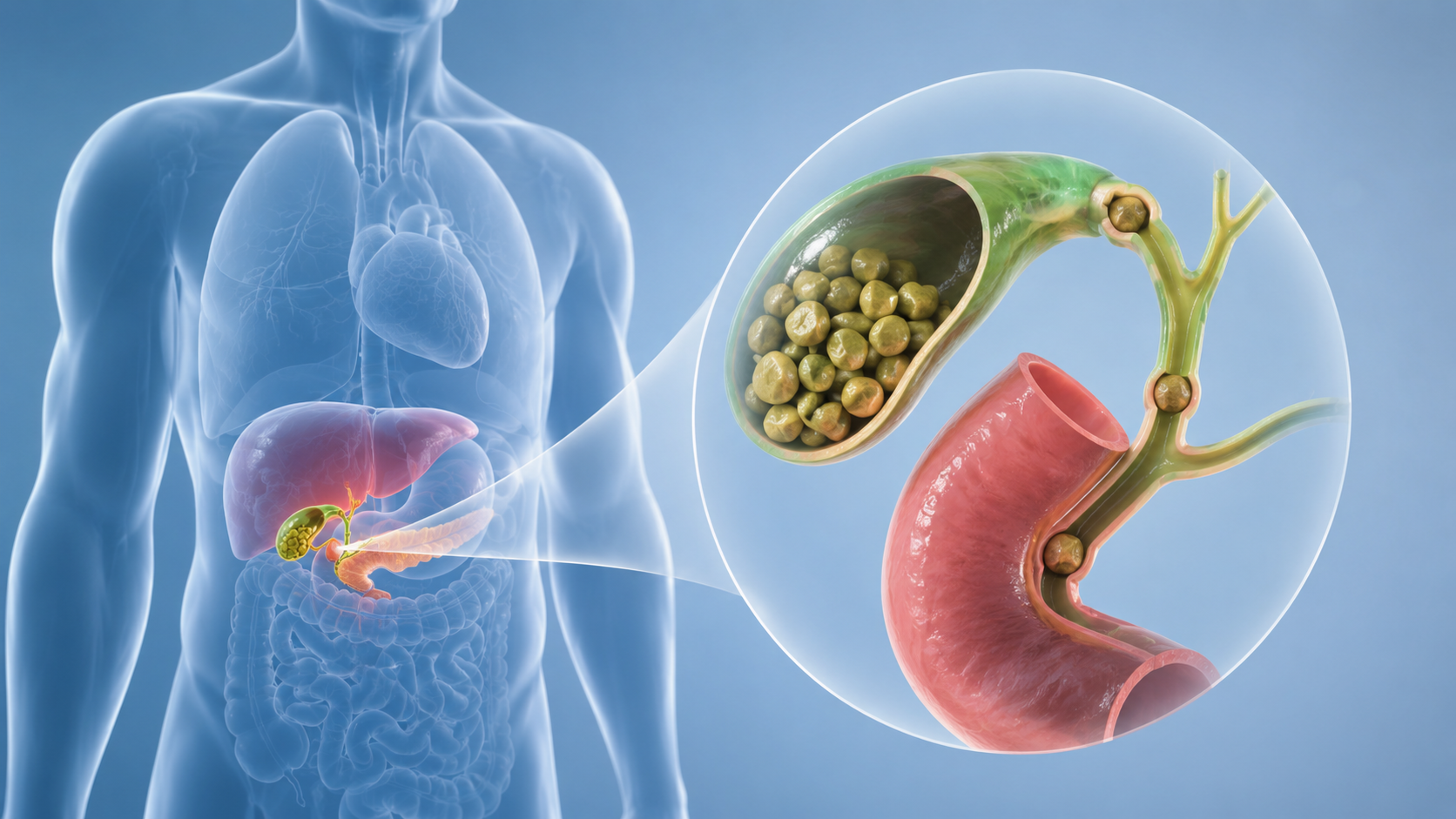
Gallbladder Stones with Common Bile Duct Stones
Gallbladder stones refer to stones located within the gallbladder. Patients may experience right upper abdominal pain, biliary colic, cholecystitis, and other symptoms.
Common bile duct stones refer to stones that have entered or formed within the common bile duct. The common bile duct acts as the “main pipeline” for bile drainage. If it becomes blocked by stones, bile cannot flow smoothly into the intestine, potentially leading to dark urine, yellowing of the skin or eyes, abnormal liver function, cholangitis, and other problems.
What Symptoms Should Raise Suspicion of Common Bile Duct Stones?
If a patient with gallbladder stones experiences the following, they should be alert to the possibility that stones have affected the common bile duct:
Right upper or upper abdominal pain, especially after eating; yellowing of the whites of the eyes or skin; abnormal liver function tests; signs of cholecystitis or cholangitis; imaging studies revealing stones in the common bile duct.
In such cases, it is advisable to consult a hepatobiliary surgeon promptly, as common bile duct stones can further lead to acute cholangitis, biliary pancreatitis, liver function impairment, and other complications.
Limitations of Traditional Treatment Approaches
In the past, the common treatment for gallbladder stones with common bile duct stones was laparoscopic cholecystectomy combined with ERCP stone extraction.
ERCP involves entering the digestive tract through the mouth and managing common bile duct stones at the duodenal papilla. This method is widely used but has some drawbacks.
First, many patients require two separate procedures: one to address the common bile duct stones and another to remove the gallbladder. This increases the treatment process, hospitalization burden, and psychological stress.
Second, ERCP stone extraction often requires sphincterotomy of the sphincter of Oddi. The sphincter of Oddi is an important structure at the opening of the bile and pancreatic ducts, regulating the flow of bile and pancreatic juice. If it is cut, its physiological function may be compromised, increasing the risk of duodenal fluid reflux, recurrent cholangitis, and stone recurrence.
Additionally, ERCP itself carries risks of complications such as pancreatitis, bleeding, and perforation.
Therefore, the ideal treatment direction is to solve the problem in a single procedure, with minimal trauma, and without disrupting the original structure and function of the biliary tract as much as possible.
Single-Port Laparoscopy Combined with Peroral Cholangioscopy
Single-port laparoscopy combined with peroral cholangioscopy is a minimally invasive treatment that integrates laparoscopic and cholangioscopic techniques.
The surgeon performs laparoscopic procedures through a small incision at the umbilicus, while simultaneously accessing the digestive tract through the mouth to enter the common bile duct and remove stones under direct visualization.
The core concept of this approach is: only one small abdominal incision, no choledochotomy, minimal injury to the sphincter of Oddi, complete clearance of common bile duct stones under direct vision, and finally management of the gallbladder.
Overview of the Surgical Procedure
The surgery is typically performed under general anesthesia.
The surgeon first makes a small incision of about 2 cm at the umbilicus, through which a single-port laparoscopic access channel is established. Because the incision is located in the umbilicus, the postoperative scar is relatively inconspicuous.
After entering the abdominal cavity, the surgeon identifies the gallbladder, cystic duct, and cystic artery under laparoscopy, and dissects the Calot’s triangle area. Then, a small incision is made on the side of the cystic duct, and a guidewire is passed through the cystic duct into the common bile duct, and further through the duodenal papilla into the intestine.
Next, the surgeon inserts a duodenoscope through the mouth to reach the duodenal papilla and locate the guidewire. Then, using a cholangioscope, the surgeon advances along the guidewire into the common bile duct, visualizes the stones under direct cholangioscopic view, and retrieves them with a basket.
After stone removal, the surgeon re-examines the bile duct to confirm no residual stones. Subsequently, the cystic duct and cystic artery are managed, the gallbladder is excised, and removed through the umbilical incision.
The characteristics of the entire process are: only one small abdominal incision, no need to open the common bile duct, and stones within the bile duct can be cleared under direct visualization.
Advantages of Single-Port Laparoscopy Combined with Peroral Cholangioscopy
- It allows for a single anesthesia and a single surgery to simultaneously address gallbladder stones and common bile duct stones. Patients do not need to undergo repeated invasive procedures in stages, making the treatment process more streamlined.
- Only one small umbilical incision is needed on the abdomen. The umbilicus has natural folds, so the incision is relatively hidden after healing, resulting in less visible trauma and better cosmetic outcomes.
- No need to incise the common bile duct. Traditional common bile duct exploration sometimes requires choledochotomy and postoperative T-tube placement. Single-port laparoscopy combined with peroral cholangioscopy can accomplish stone removal through the cystic duct and the natural orifice of the digestive tract, avoiding direct choledochotomy as much as possible.
- It can avoid T-tube placement. T-tubes bring postoperative care inconvenience and may affect the patient’s quality of life. Being able to “avoid T-tube placement” is an important advantage for postoperative recovery.
- It preserves the function of the sphincter of Oddi as much as possible. Compared with sphincterotomy, this technique emphasizes protecting the normal structure of the biliary outlet, reducing disruption to the physiological function of the biliary tract.
- Stone removal can be performed under direct visualization. The cholangioscope allows direct observation of the interior of the common bile duct, showing the location, number, and residual status of stones, which helps improve the thoroughness of stone clearance and reduce the chance of missed stones.
The Importance of Direct-Vision Stone Removal
The key to treating common bile duct stones is to ensure, as much as possible, that all stones have been removed.
If reliance is placed solely on indirect assessment, there may be a risk of residual stones. The cholangioscope can enter the common bile duct for internal observation, transforming the previous blind extraction into removal under direct vision. The surgeon can more clearly see whether stones have been completely cleared and can further examine the condition inside the bile duct.
This is particularly important for patients with multiple stones, impacted stones, or complex common bile duct conditions.
Avoiding Choledochotomy
The common bile duct is an important channel for bile drainage. Although choledochotomy can remove stones, it may also lead to problems such as bile leakage, stricture, infection, and the need for postoperative T-tube placement.
If stone removal can be accomplished through the cystic duct and peroral cholangioscopy, direct trauma to the common bile duct itself can be reduced.
Preserving the Sphincter of Oddi
The sphincter of Oddi is located at the exit of the bile and pancreatic ducts into the duodenum. It participates in regulating the flow of bile and pancreatic juice, and also serves to prevent intestinal fluid reflux into the biliary tract.
If this structure is cut, the normal barrier function of the biliary tract may be compromised. In the long term, it may increase problems such as duodenal fluid reflux, recurrent cholangitis, and recurrence of common bile duct stones.
Which Patients Are Suitable?
The single-port laparoscopy combined with peroral cholangioscopy technique is mainly suitable for patients with gallbladder stones and common bile duct stones, especially those who wish to address stones in both locations in a single surgery and avoid choledochotomy, T-tube placement, and injury to the sphincter of Oddi as much as possible.
However, suitability must be determined based on the specific condition, including stone size, number, and location, common bile duct diameter, cystic duct anatomy, presence of acute inflammation, liver function status, presence of bile duct dilation, and the operating team’s experience.
Does It Require High Technical Skill from the Surgeon?
The single-port laparoscopy combined with peroral cholangioscopy technique demands a relatively high level of technical skill from the surgeon.
Single-port laparoscopy itself has limited operating space, with all instruments entering through a single umbilical channel, which can easily interfere with each other. Peroral cholangioscopy also requires coordination with the endoscopy team and the ability to accurately enter the common bile duct under guidewire guidance and complete stone removal.
Therefore, combined gallbladder and bile duct stone removal requires collaboration among a team of physicians with expertise in hepatobiliary surgery, endoscopic techniques, and cholangioscopy operation.